CASE
HISTORY / CLINIC:
55-year-old patient with no symptom of pain. Alcohol abuse and consumption of
30 cigarettes per day for several years. A hard and painful area with pains that
radiate into the ear was palpated around the left tongue base during the otorhinolaryngological
examination.
FINDINGS:
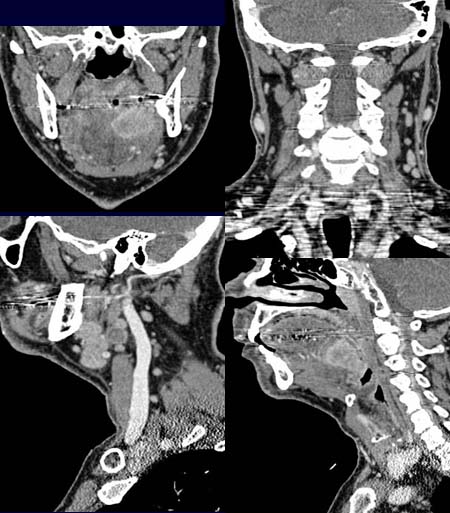
The coronal CT scan after contrast administration (upper left image) reveals
a contrast-enhancing
mass lesion on the left side of the tongue base/floor of the mouth that extends
right up to the midline.
The coronal and sagittal reformation images (upper right and lower left images)
demonstrate multiple
pathologic lymph nodes.
The craniocaudal
extension of the tumor is well demonstrated in the sagittal, left paramedian
reformation image (lower right image).
DIAGNOSIS:
Oropharynx carcinoma
DIFFERENTIAL DIAGNOSIS:
Other malignant masses of the oral cavity.
Web resources: