CASE
HISTORY / CLINIC:
Heavy pipe smoker. Reddened, slightly ulcerated area in the anterior portion
of the mouth floor for some weeks. Postprandial swelling in the left submandibular
gland that consistently persists after glandular massage.
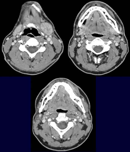
FINDINGS:
The left submandibular gland appears swollen
and congested in the soft-tissue window scan (upper left picture). The
right submandibular gland appears normal.
The congested
excretory duct of the left submandibular gland can be seen in the CT scan
at a more superior position (upper right picture). A small hyperdense
mass is seen in the CT scan at an even more superior position (lower image).
DIAGNOSIS:
Small carcinoma in the anterior portion of the mouth floor with congestion
of the submandibular gland.
DIFFERENTIAL DIAGNOSIS:
Other tumors of the mouth floor.
Web resources:
1) Medecoinfo