CASE
HISTORY / CLINIC:
Patient who had undergone radio-chemotherapy of a lymphoma presenting with partially
purulent nasal discharge. Patient coughs out partialy
bloody crusts. The mirror examination revealed white-yellowish fibrin-coated
areas in the nasopharyx.
FINDINGS:
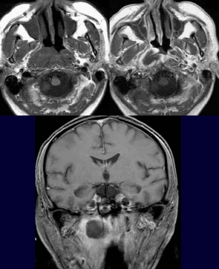
The non-contrast T1-weighted axial image (left image above)
demonstrates a large,
mainly right-sided nasopharyngeal mass. The mass shows a rim
enhancement and central hypodensity in the post-contrast T1-weighted axial
images (right image above).
The mass is seen mainly in the right nasopharynx and has
a hypodense
center and a hyperdense
(contrast-enhancing) periphery in the post-contrast
T1-weighted coronal images (picture below)
DIAGNOSIS:
Aspergillose
DIFFERENTIAL DIAGNOSIS:
Lymphom oder Malignom des Nasopharynx
Web resources: