CASE
HISTORY / CLINIC:
Patient presenting with emergent progressive loss of visual acuity and double
images. The endoscopic examination of the head and neck region revealed no pathological
findings.
FINDINGS:
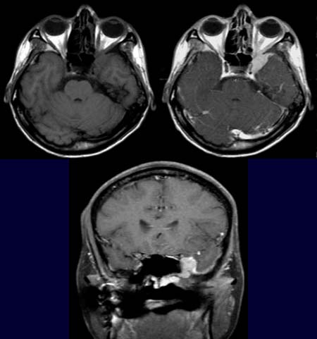
The T1-weighted axial image reveals a hypodense
mass around the left side of the sphenoid bone, left orbita and left temporal
lobe. The mass is seen to enhance intensely in the T1-weighted axial image after
contrast administration (upper left picture). Contrast enhancement is also demonstrated
around the left carotid siphon. A definite demarcation from left the orbita is not
posible.
The mass beside the
ethmoid sinus at the skull base appears hyperdense in
the T1-weighted coronal images
done after contrast administration. The image further
demonstrates an enhancement
of the dura on the temporal side, most probably due to a tumorous infiltration.
DIAGNOSIS:
Adenoid cystic carcinoma
DIFFERENTIAL DIAGNOSIS:
Meningeoma or other malignant tumor of the skull base
Web resources: