CASE
HISTORY / CLINIC:
25-year patient with severely impaired nasal breathing, increasing headache and
incipient loss of visual acuity on the right side.
The nasal endoscopy revealed a round well defined reddish tumor.
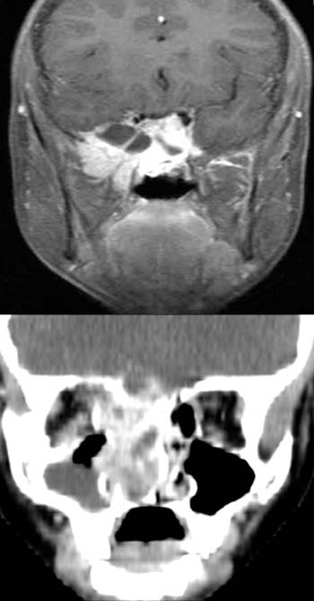
FINDINGS:
The fat suppression, contrast-enhanced T1 MRT image (upper picture) demonstrates
an intensely
enhancing mass with areas
of low signal in the sphenoid sinus and right pterygopalatine fossa. The
mass extends bilaterally (although to a greater extent on the right side) to
the temporal
lobes. The mass borders on the internal
carotid artery both sides around the sphenoid sinus.
The coronal CT (lower picture) also demonstrates a large,
strongly enhancing mass in the main nasal cavity and ethmoidal sinus (mainly
the right side). The mass extends to the orbital cone and seems to spread intracranially
around the lamina cribrosa. The right
maxillary sinus partially appears radio-opaque suggesting fluid collection.
DIAGNOSIS:
Large nasopharyngeal fibroma
DIFFERENTIAL DIAGNOSIS:
Malignant tumor in the paranasal sinuses and main nasal cavity.
Suggestions for further reading: