CASE
HISTORY/CLINIC:
38-year-old female with loss of visual
acuity on the right eye.
FINDINGS:
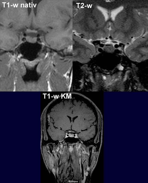
The T2-w image shows evidence of a dull hyperintense spotty lesion of
the optic nerve on the right– in comparison with the left optic nerve the
right optic nerve shows a discrete swelling that
is also seen on T1-w. The postcontrast T1-w shows spotty contrast
enhancement of the right optic nerve.
DIAGNOSIS:
Optic
neuritis associated with disseminated
encephalitis
DIFFERENTIAL DIAGNOSIS:
Optic glioma
web
resources:
1) ACR Learning file
2) AWMF
3) Medicine worldwide
4) MRI-Library