CASE
HISTORY/CLINIC:
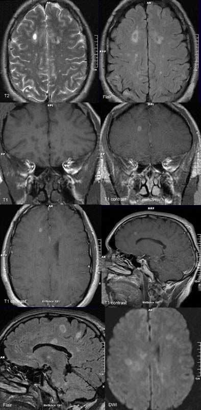
43-year-old female with paresthesia.
FINDINGS:
The T2-w and FLAIR images show evidence of multiple oval, periventrikular lesions
in the medullary layer as well as in the corpus
callosum and pons.
The above mentioned lesions appear hypointense on
T1-w (black holes) and show dull spotty
or ring-shaped enhancement
after contrast administration
DIAGNOSIS:
Demyelinating
foci of disseminated encephalitis that are partially
enhancing, just like in an acute episode.
DIFFERENTIAL DIAGNOSIS:
Vascular lesions
ADEM (acute disseminated encephalomyelitis)
Metastases
Multiple abscesses
Toxoplasmosis
web
resources:
1) ACR Learning file
2) AWMF
3) Medicine worldwide
4) MRI-Library