CASE
HISTORY/CLINIC:
36-year-old female with diagnosed malignant melanoma on the left lower leg.
FINDINGS:
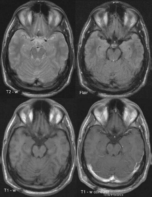
Hyperintense right-sided parietooccipital subcutaneous mass
lesion on
T2-w and FLAIR images with prominent enhancement on
postcontrast T1.
Due to the high melanin content the mass lesion shows a high
signal even
on the precontrast T1 image.
DIAGNOSIS:
Mainly subcutaneous melanoma
metastasis or lymph node metastasis.