CASE
HISTORY/CLINIC:
58-year-old patient with head injury sustained about 3 months ago.
FINDINGS:
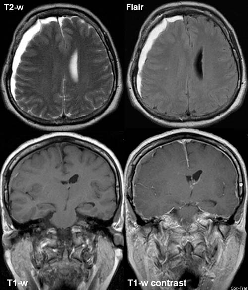
The T2-w and flair images show evidence of a homogeneous high
signal sickel-shaped right-sided frontoparietal
and left-sided frontal
parafalcine structure.
The lesion exceeds the cranial sutures and causes compression of
the right lateral ventricle and mid-line shift to the left.
The fluid
collection in the pre-
and postcontrast T1-w is homogeneous and has a signal intensity similar
to that of the gray matter and shows no contrast enhancement. Contrast
enhancement of the Meninges.
DIAGNOSIS:
Right-sided frontoparietal as
well as left-sided parafalcine subdural
hematoma resulting in mid-line
shift to the left and compression of the right lateral
ventricle. Meningeal
stimulation.
web
resources:
1) ACR Learning file
2) MRI-Library