CASE
HISTORY/CLINIC:
14-year-old patient after a head injury about 15 days ago.
FINDINGS:
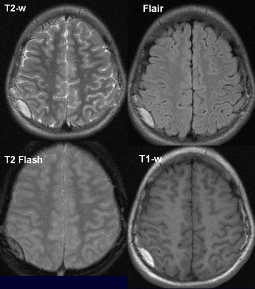
The T2-w, Flair und T1-w show evidence of homogeneous, hyperintense, convex-shaped dorsoparietal mass
lesion on the right. The lesion doesn't exceed the cranial sutures.
The structure is surrounded by a soft peripheral
margin with a low signal.
A reduction
in signal intensity, the so-called Subzibilitätsartefakten caused
by the magnetic effekt of blood degradation products is evident on the T2-Flash
image.
The lesion shows no contrast
enhancement in the postcontrast T1-w (not visualized here).
DIAGNOSIS:
Subacute
dorsoparietal hematoma on the right.
DIFFERENTIAL DIAGNOSIS:
Small subdural hematoma
web
resources:
1) MRI-Library