CASE
HISTORY
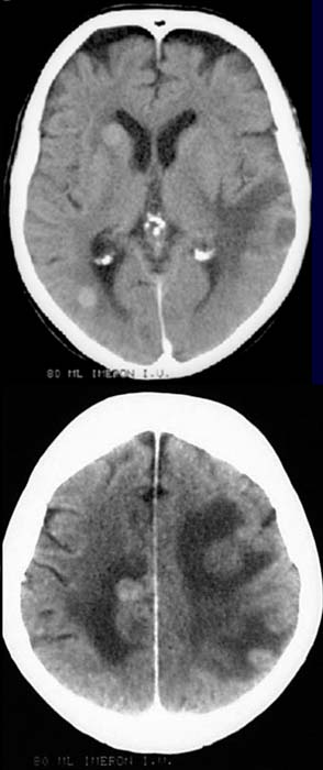
/ CLINIC:
54 years old patient with diagnosedjähriger bronchial carcinoma. Severe
headache.
FINDINGS:
CCT after contrast admistration.
Multiple
hyperdense contrast-enhancing mass
lesions on the left temporal lobe, right frontal lobe, right basal ganglia,
right occipital lobe and the upper part of both parietal lobes . Hypodense
area surrounding the mass lesions.
DIAGNOSIS:
Multiple
cerebral metastases at diagnosed bronchial carcinoma with perifocal
oedemas.
Information on the internet:
1) Uni Harvard
2) Neuropat