CASE
HISTORY:
50-year-old patient, unconscious
after a severe craniocerebral trauma.
FINDINGS:
The unenhanced
control CT-scan (i.e.
without contrast administration)
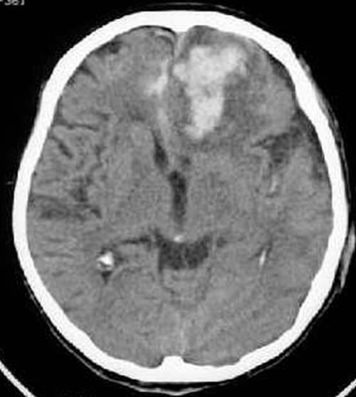
after about a week shows: Hyperdense (60-80HE) bifrontal
intracerebral mass
(predominantly on the left) with surrounding hypodense (20-30 HE) rim.
Hyperdense
areas between the hemispheres. Asymmetric
appearance of the ventricular system. Slight frontal midline
shift to the right.
DIAGNOSIS:
Not quite fresh left-sided frontal contusion
hemorrhage, slightly also on the right. Perifocal
edema and subarachnoid
hemorrhage.
DIFFERENTIAL DIAGNOSIS:
Hemorrhagic
insult
Tumor
Abscess