CASE
HISTORY:
A
71-year-old patient with a history of hypertension suddenly suffers from severe
headache and imbalance. Patient is comatose.
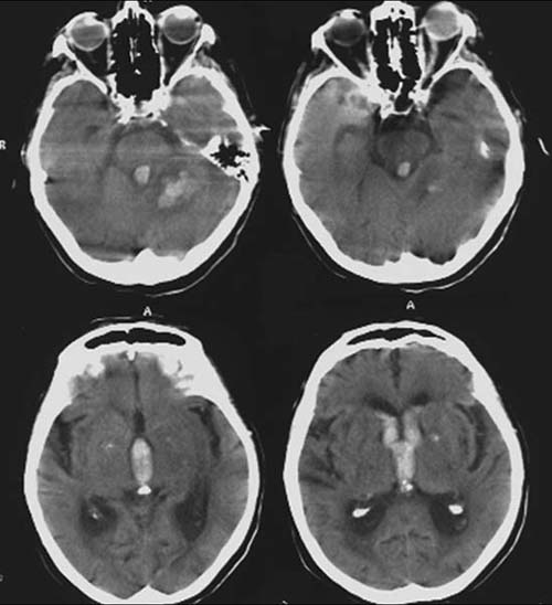
FINDINGS:
Unenhanced CCT-scan (i.e.
without contrast administration).
Hyperdense
(60-80HE) mass in the left cerebellar hemisphere with perifocal
hyperdensity. Hyperdense
collection in the entire ventricular system. Flattened
gyri and sulci of the left cerebellar hemisphere. Hyperdensity
of the corpus vitreum of the left eye in comparison to the right eye.
DIAGNOSIS:
Massive
hypertensive
hemorrhage of the left cerebellar hemisphere and perifocal
edema.
Hemorrhage
into the ventricle system.
Edema
of especially the left cerebellar tonsils.
Intravitreal
hemorrhage
DIFFERENTIAL DIAGNOSIS:
Brain
tumors (glioblastoma, metastases)
Hemorrhagic infarction
Cerebral abscess