CASE
HISTORY:
A
71-year-old patient with a history of hypertension suddenly suffers from severe
haedache and imbalance. Patient is comatose.
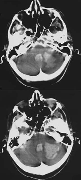
FINDINGS:
Unenhanced
CCT (i.e.
without contrast administration).
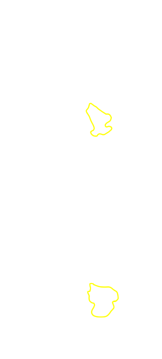
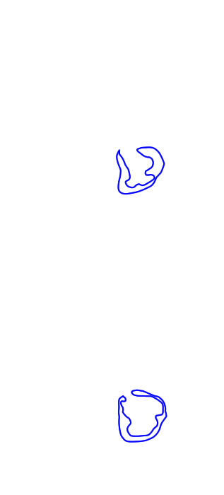
Hyperdense
(60-80HE) mass in the left cerebellar hemisphere with perifocal
hyperdensity. Hyperdense
filling in the the fourth ventricle. Flattened
gyri und sulci of the left cerebellar hemisphere.
DIAGNOSIS:
Massive
hypertensive
hemorrhage of the left cerebellar hemisphere and perifocal
edema.
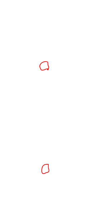
Hemorrhage
into the fourth ventricle.
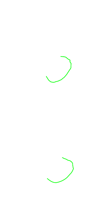
Edema
of especially the left cerebellar tonsils.
DIFFERENTIAL DIAGNOSIS:
Brain
tumors (glioblastoma, metastases)
Hemorrhagic infarction
Cerebral abscess