CASE
HISTORY / CLINIC:
Patient with reddish-bluish lesion around the left lips and cheeks since birth.
Lesion has been gradually progressing
for a few years now and is soft and non-dolorous upon palpation.
FINDINGS:
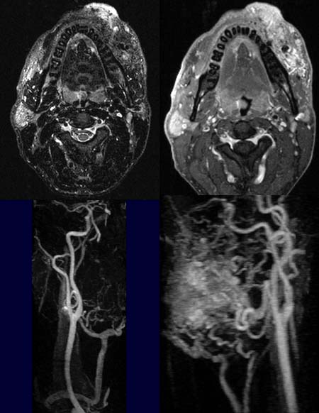
Demonstration
of a large inhomogeneous
mass lesion in the left cheek and on the left side of the lip as seen
in the T2 image (upper left image). Signal
effacement is seen, typical of calcifications.
The margins
of the mass are even better seen in the contrast-enhanced T1 image (upper
right image). Signal
effacements are also seen here in association with thrombosed vessels or
calcifications.
The right side appears normal in the MR angiography (lower left image).
The MR angiography of the left side (lower right image) demonstrates the extensive
vascularization of the tumor by branches of the external carotid artery.
DIAGNOSIS:
Hemangioma
of the cheek and lips
DIFFERENTIAL
DIAGNOSIS:
Lymphangioma
Web
resources: