CASE
HISTORY / CLINIC:
55-year-old patient with increasing swelling in the entire upper jaw. Double
images and severe pains for 4 weeks now. Particularly hypesthesia around the infraorbital
nerve on both sides.
FINDINGS:
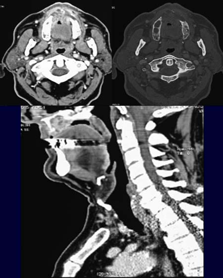
The
axial scan of the upper jaw (upper left image) demonstrates a large mass
lesion. This mass lesion has eroded the alveolar bone of the upper jaw ventrally. The
erosion is best seen in the bony window image (upper right image).
The anteroposterior
extension is well seen in the sagittal reformation image (lower image).
DIAGNOSIS:
Maxillar
carcinoma
DIFFERENTIAL
DIAGNOSIS:
Other
malignant tumors of the mouth floor.
Web
resources:
1) Medecoinfo