CASE
HISTORY / CLINIC:
51-year-old heavy smoker with increasing pains in the lower jaw.
FINDINGS:
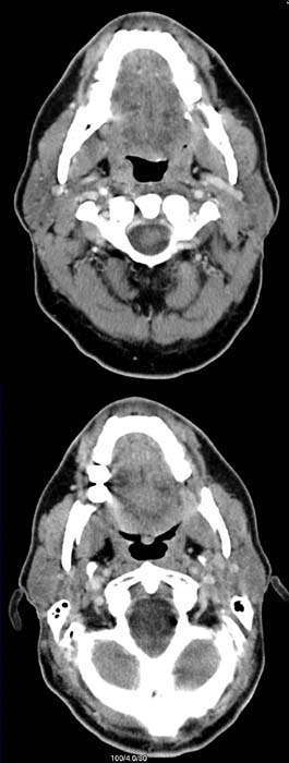
The contrast-enhanced axial image of the oral cavity (upper image) demonstrates
a contrast-enhancing
mass in the left angle of the jaw (region 38). There was an 80 seconds delay
after the start of contrast injection. The tumor is
even better demonstrated in the delayed image of the same region after about
100 seconds (lower image).
DIAGNOSIS:
Squamous cell carcinoma in the left angle of the mandible.
DIFFERENTIAL DIAGNOSIS:
Other malignant tumors of the mouth floor.
Web resources:
1) Amersham Health
2) Medecoinfo