CASE
HISTORY / CLINIC:
Patient with postprandial swelling in the left submandibular gland. The swelling
has been recurring for two years.
Patient currently feels no pain and has no other complaints.
FINDINGS:
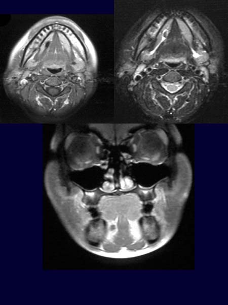
The post contrast T1-weighted image (upper left picture) depicts a poorly
enhanced area in the floor of the mouth around the excretory duct of the
right submandibular gland. The excretory
duct of the left submandibular gland appears somewhat
enlarged in the T2-weighted
axial image (upper right picture). An
area of low signal intensity can be seen in front of this enlargement.
The post contrast coronal T1-weighted sequence demonstrates an enlarged
duct of the right submandibular gland (lower picture).
DIAGNOSIS:
Sialolithiasis
DIFFERENTIAL DIAGNOSIS:
Mass lesion in the anterior floor of the mouth.
Web resources:
1) Amersham Health
2) Medecoinfo
3) Pathologie Online
4) MRX