CASE
HISTORY / CLINIC:
67-year-old patient with hoarseness and severe dysphagia.
FINDING:
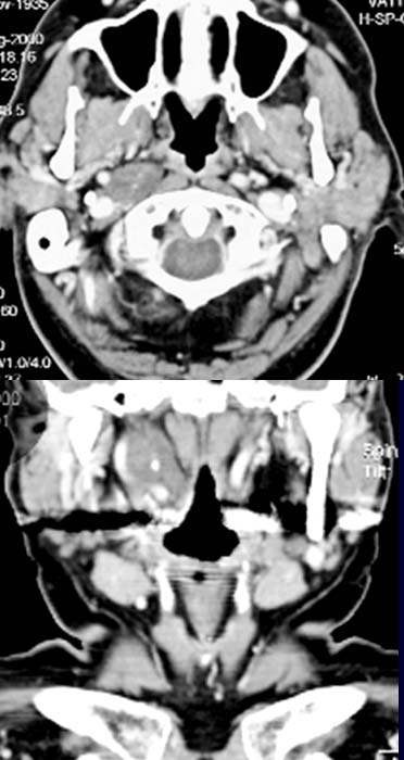
The axial CT image (upper picture) demonstrates a well
defined hyperintense homogeneous mass measuring
about 2 x 1.5 cm in diameter in the right parapharyngeal space. The neighboring
structures, especially the blood
vessels are distincly demarcated from the mass. The coronal reconstruction
image (lower picture) clearly demonstrates the cranio-kaudal dimensions of the tumor likewise
the lateral displacement of the internal
carotid artery without any evidence of invasion.
DIAGNOSIS:
Neurinoma of the vagus nerve.
DIFFERENTIAL DIAGNOSIS:
Neurinoma of a different nerve.