CASE
HISTORY / CLINIC:
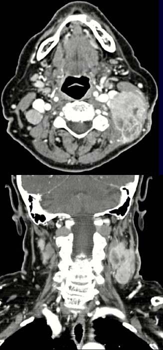
55-year-old patient with a known history of hypopharyngeal squamous cell carcinoma.
FINDING:
The post contrast CT image shows a large hyperdense mass below
the left sternocleidomastoid with
typical central
necrolysis. Another mass 2
cm in diameter showing no signs of central necrolysis is also demonstrated behind
the large vessels on the other side. A small right-sided
submandibular mass without central necrolysis is also demonstrated.
The coronal image (lower picture) clearly demonstrates the cranio-caudal spread
of these masses on
both sides of the parapharyngeal space.
DIAGNOSIS:
Metastases of a squamous
cell carcinoma on both sides
DIFFERENTIAL
DIAGNOSIS:
Other lymph node metastases
Web resources: