CASE
HISTORY / CLINIC:
Patient with a swelling in the left parotid gland.
The swelling is painless and relatively homogeneously palpable. Night sweat and
weight loss. The
facial nerve is not affected.
FINDINGS:
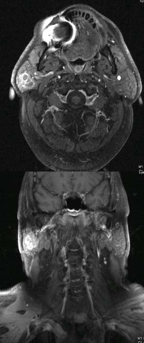
The
post-contrast T1-weighted axial image (above) demonstrates a multicystic,
relatively well defined tumor. A signal
interference can be seen as a side finding in the lower jaw, corresponding
to a distinctive metal artifact of a dental prosthesis (red)
The post-contrast T1-weighted coronal image shows the cranio-caudal spread of
the tumor. The image also depicts a multicystic,
relatively well circumscribed, hyperdense tumor.
DIAGNOSIS:
Mukoepidermoid carcinoma.
DIFFERENTIAL
DIAGNOSIS:
Other
malignant tumors of the parotid gland.
Web
resources:
1) Amersham Health
2) Medecoinfo
3) Roche Lexikon Medizin
4) Pathologie Online