CASE
HISTORY / CLINIC:
Patient with pronounced hinderance of nasal breathing. Condition has been progressive
for many months now. Swollen lymph nodes on both sides of the neck a couple of
weeks ago.
FINDINGS:
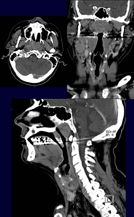
The axial CT image after contrast administration (upper left picture) reveals
a nearly total occlusion of the nasopharynx by a poorly
enhancing mass. The mass appears a
bit hyperdense, occluding the nasopharynx in the coronal image after contrast
administration (upper right image). The ethmoid sinus that lies superioir to
the mass is clear.
The sagital image after contrast administration excellently demonstrates the cranio-caudal
spread of the nasopharyngeal tumor
as well as the demarcation
from the clivus.
DIAGNOSIS:
Nasopharyngeal carcinoma
DIFFERENTIAL DIAGNOSIS:
Lymphoma
Web resources:
1) Amersham Health
3) Roche Lexikon Medizin
3) Medicine Worldwide
4) Pathologie Online