CASE
HISTORY / CLINIC:
Patient from Hongkong with recurring episodes of middle ear effusions on the
left.
The mirror examination of the nasopharynx revealed hyperplastic lymphatic tissue
in the mucous membrane on the left side.
FINDINGS:
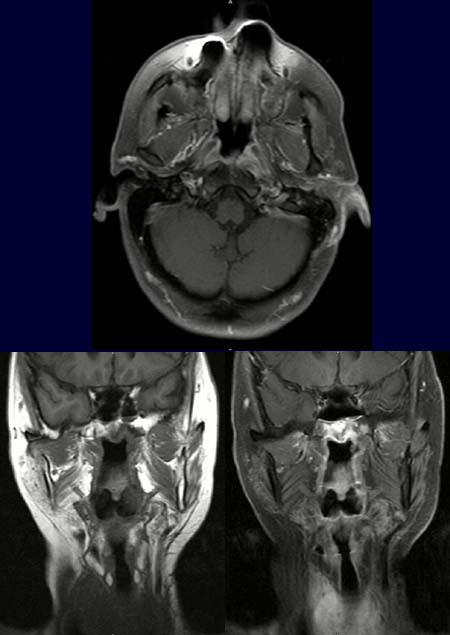
The T1-weighted axial image
of the nasopharynx after contrast administration (upper
image) reveals a hyperdense
mass around the origin of the left eustachian tube. The pre-contrast T1-weighted
coronal image (lower left picture) reveals an assymetry in the nasopharynx with large
hypodense area on the left side. The tumor
appears hyperdense in the T1-weighted coronal image after contrast administration
(lower right image).
DIAGNOSIS:
Nasopharyngeal carcinoma.
DIFFERENTIAL DIAGNOSIS:
Lymphoma
Hyperplastic lymphatic tissue
Web resources:
1) Amersham Health
3) Roche Lexikon Medizin
3) Medicine Worldwide
4) Pathologie Online