CASE
HISTORY / CLINIC:
45-year-old patient with muffled voice.
FINDINGS:
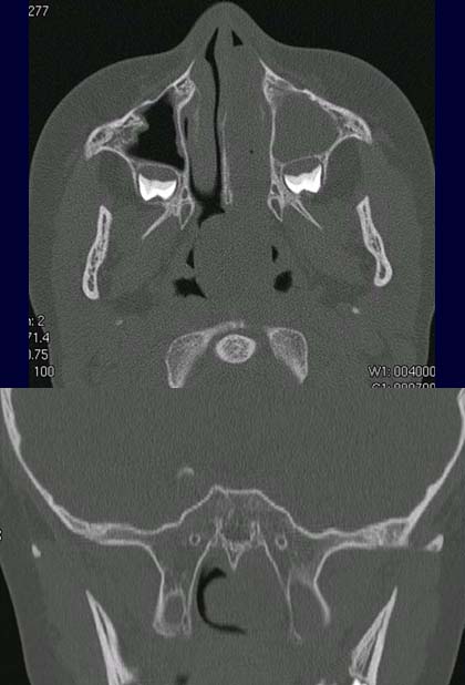
The
axial CT scan at bony window (upper image) demonstrates a mass in
both the left and right nasal cavities and in the nasopharynx. The mass obstructs
the drainage of the left maxillary sinus thus causing collection
of secretion .
Secondary finding: dental
germs behind both maxillary sinuses. The mass is
viewed between the pterygoid
processes in the coronal scan at bony window (lower picture); it fills up
the whole nasopharynx.
DIAGNOSIS:
Choanal
polyp
DIFFERENTIAL
DIAGNOSIS:
Inverted
papilloma, Lymphoma
Suggestions
for further reading: