CASE
HISTORY / CLINIC:
57-year-old man with increasing nasal obstruction for months. Left-sided exophthalmus
and increasing protrusion in the left cheek for some weeks now.
FINDINGS:
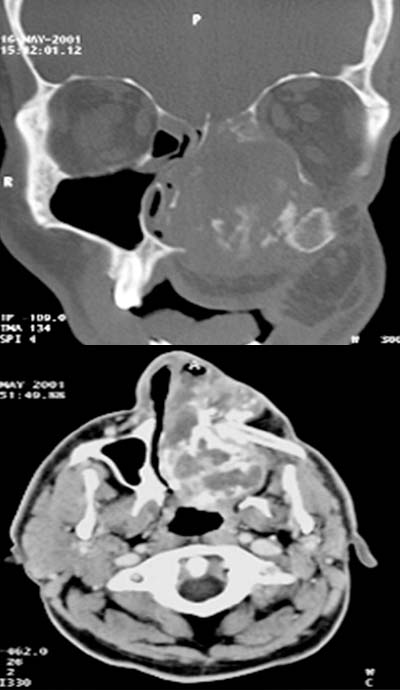
The coronal CT scan at bony
window demonstrates a large
mass lesion in the left nasal cavity and ethmoidal cells. The nasal septum
is displaced to the right. The hard
palate is missing due to a previous operation. The mass demonstrates laminar calcifications and extends
cranially into the left orbital cone. The axial CT scan also demonstrates
this tumor with laminar calcifications.
A fibula
graft is visualized in the location of the left maxilla due to the previous
operation.
DIAGNOSIS:
Large relapse of an osteosarcoma located in the left nasal cavity and extending
to the palate and left orbita.
DIFFERENTIAL DIAGNOSIS:
Chondrosarcoma
Suggestions for further reading:
1) Amersham Health
2) Medecoinfo
3) Medicine Worldwide
4) AHC-Consilium