CASE
HISTORY / CLINIC:
78-year-old patient with purulent discharge from the left nose. Partly pressure
pains in the orbita, and over the frontal and maxillary sinuses. A reddish/bluish
tumor occupying the entire left nasal cavity and which easily bleeds when touched
was found during endoscopy.
FINDINGS:
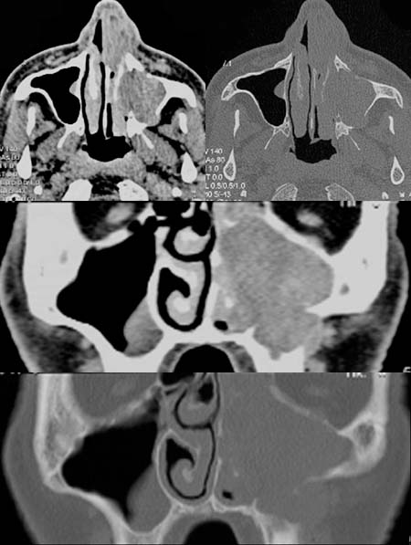
The
contrast-enhanced axial CT scans at soft-tissue and bony windows demonstrate
a hyperdense,
contrast-enhancing mass lesion in the anterior portion of the nasal cavity
and in the left maxillary sinus. The wall of the maxillary sinus is laterally
discontinuous, the mass extends into the masticatory space. The extension
beyond the lateral wall of the maxillary sinus is best viewed in the
coronal reformation images(pictures at the bottom).
DIAGNOSIS:
Malignant
melanoma of the left nasal cavity and left maxillary sinus.
DIFFERENTIAL
DIAGNOSIS:
Other
malignant tumors of the paranasal sinuses.
Suggestions
for further reading:
1) Amersham Health
2) Medecoinfo
3) Medicine Worldwide
4) Onkologie.de