CASE
HISTORY / CLINIC:
66-year-old female coming for a follow-up examination after an operation of a
cranial base tumor ten years ago. Granulating tissue was found in the right frontal
sinus during the endoscopic examination performed.
FINDINGS:
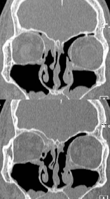
The coronal CT scan (bony window) without contrast administration demonstrates
a hypodense
mass in the ethmoidal cells and frontal sinus. The superior
nasal concha on the left side and the walls of the ethmoidal cells are missing
due to the previous operation.
Side finding: polypoid
swelling of the mucous membrane lining both maxillary sinuses.
DIAGNOSIS:
Relapse of an esthesioneuroblastoma after
a previous operation. No evidence of intracranial orbital spread on the right side
.
DIFFERENTIAL DIAGNOSIS:
Other malignant or benign tumors in the ethmoidal cells and frontal sinuses.
Suggestions for further
reading:
1) Amersham Health
3) AWMF