CASE
HISTORY / CLINIC:
52
year old heavy smoker with severe swallowing difficulties.
FINDINGS:
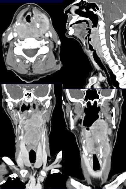
The post
contrast axial CT image of the larynx demonstrates an extensive, mainly left-sided mass on
both sides of the larynx with distinct inhomogeneous contrast enhancement.
The mass can be seen all around laryngeal
skeleton. The lumen of the larynx is slightly displaced to the left.
The sagittal reconstruction image (top right image) excellently demonstrates
the cranio-caudal spread of the tumor that
extends from the oropharynx right down to the larynx. The coronal reconstruction
images (images below)
also demonstrate the spread of the tumor;
the lower right picture
shows the growth all around of the laryngeal skeleton. The lower left picture
also shows lymph
node metastases.
DIAGNOSIS:
Extensive hypopharynx-larynx carcinoma with pathological lymph nodes.
DIFFERENTIAL DIAGNOSIS:
Other
malignant tumors with origin in the hypopharynx or larynx.
Web
resources:
1) Amersham Health
2) Roche Lexikon Medizin
3) Medicine Worldwide
4) Pathologie Online
5) Onkologie.de
6) MRX.de