CASE
HISTORY / CLINIC:
56-year-old patient with severe dysphagia and incipient shortness of breath.
FINDINGS:
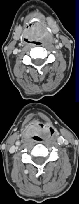
Demonstration of an extensive, mainly left-sided contrast-enhancing mass on
both sides of the epiglottis at the level of the hyoid.
The mass seems to be confined to the epiglottis and only borders ventrally on
the hyoid and dorsally on the posterior wall of the hypopharynx. The sheath
of nerves and vessels has not been invaded. In the CT image done at a lower
level after contrast administration (right) the epiglottic mass shows
an inhomogeneous contrast enhancement.
Both images demonstrate some lymph
nodes in the sheath of nerves and vessels on the right
side.
DIAGNOSIS:
Leiomyosarcoma of the epiglottis without evidence of pathological lymph nodes.
DIFFERENTIAL DIAGNOSIS:
Supraglottic squamous cell carcinoma