CASE
HISTORY / CLINIC:
39-year-old patient with sudden paresis of the left facial nerve. The branches
to the mouth, eye and forehead are affected. Light pains around the left ear.
Normal findings of ear microscopy .
FINDINGS:
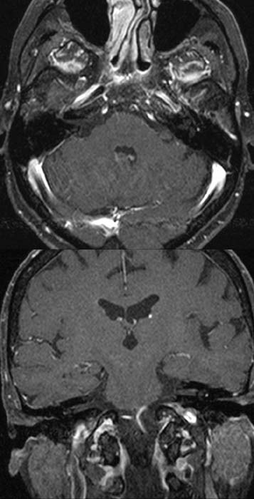
The post-contrast T1-weighted axial image of the cerebellopontine angle shows high
signal intensity along the course of the facial nerve through the left middle
ear (image above). The right facial nerve doesn't show any
hyperdensity. The post-contrast T1-weighted coronal image (picture below)
also shows a significant
enhancement of left facial nerve in it's course through the middle ear. The
right facial nerve in comparison doesn't demonstrate any
hyperdensity.
DIAGNOSIS:
Neuritis of the facial nerve most probably resulting from viral infection.
DIFFERENTIAL DIAGNOSIS:
Alteration of the facial nerve e.g due to trauma
Web resources:
1) Gesundheit.de
2) Amersham Health
3) Neuritis.de