CASE
HISTORY / CLINIC:
Patient with acute hearing loss on the right as well as pronounced rotatory vertigo
and nystagmus to the right. Normal findings of ear
microscopy.
FINDINGS:
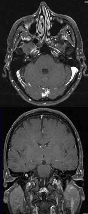
The post-contrast T1-weighted axial image through the cerebellopontine angle
(above) depicts the high signal intensity of the right cochlea as
well as parts of the right labyrinth.
The findings
in the cochlea are better seen in the post-contrast T1-weighted coronal
image (below).
DIAGNOSIS:
Cochlitis and labyrintitis most probably due to a viral infection.
DIFFERENTIAL DIAGNOSIS:
Inflammation of the inner
ear structures resulting from other diseases.