<<<
Compare
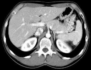
the pathological image-left and the physiological image-right
(blinded)
<<
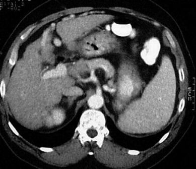
F:
Irregular
shape
of the liver . Notize
the contrast-enhanced vessels
inside the falciform ligament and near the umbilicus. Splenomegaly
H:
Adult
man, 51-years-old, admitted with reduced liver function. Alcohol abuse since
many years.
INFO/WWW-LINKS:
The most common cause of cirrhosis is alcoholic liver disease. Others are
hepatitis. Typical features of cirrhosis are varices, ascites and splenomegaly.
Esophageal varices may be caused by increased portal
venous hypertension, e.g. in cirrhosis of the liver. The elevated venous
pressure causes portosystemic shunts - like esophageal varices - to carry
portal blood into the systemic circulation. This may cause encephalopathy
and haemorrhage - especially esophageal vairces. Another portosystemic shunt
is shown in the images above: Recanalisation of the umbilical vein. Further
evidence for liver cirrhosis is an enlarged caudate lobe. The surface of the
liver has a nodular apperance, accounting for the term "hob-nail liver".
D:
Liver cirrhosis
with recanalisation of the umbilical vein
IN
THIS PART OF THE PAGE YOU FIND SOME TEXT FIELDS WHICH CAN BE OPENED EIGTHER
STEP BY STEP (CLICK ON "HISTORY", "HELP", "FINDINGS",
"DIAGNOSIS" OR "INFO/WWW-LINKS") OR AT ONCE WITH A CLICK
ON "ALL ON" - VICE VERSA CLICK ON "ALL OFF".
It is not
easy to find an exactly corresponding slice to every pathological example!
For that reason the
FILM
(2)
is
recommended!
Once opened you may use it for every pathological example.
If you need a physiological
image to compare click here