INFO/WWW-LINKS: Fresh blood is high density compared with brain on CT and, at brain windows, appears white. As the clot absorbs, the lesion becomes isodens (2-3 weeks after bleeding) and, later, of low attenuation Sites of intracranial heamorrhage/haematoma: 1) Between inner tabel and dura = extradural heamorrhage/haematoma 2) Between dura and arachnoid = subdural heamorrhage/haematoma 3) Between arachnoid and brain surface = subarachnoid heamorrhage/haematoma 4) Within brain = intracerebral heamorrhage/haematoma 5) Within ventricles = intraventricular heamorrhage/haematoma Rising intracranial pressure can be caused by e. g. head injury, meningoencephalitis, haemorrhage or cerebral oedema and tumour
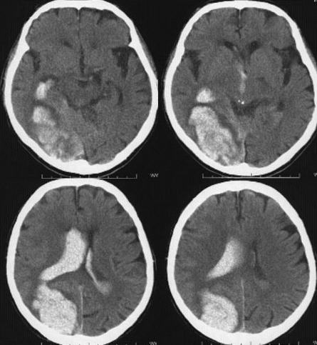
D: 1) Acute right-sided intracerebral occipital haemorrhage and haemorrhage into the ventricular system (especially the right lateral ventricle).