CASE
HISTORY/CLINIC:
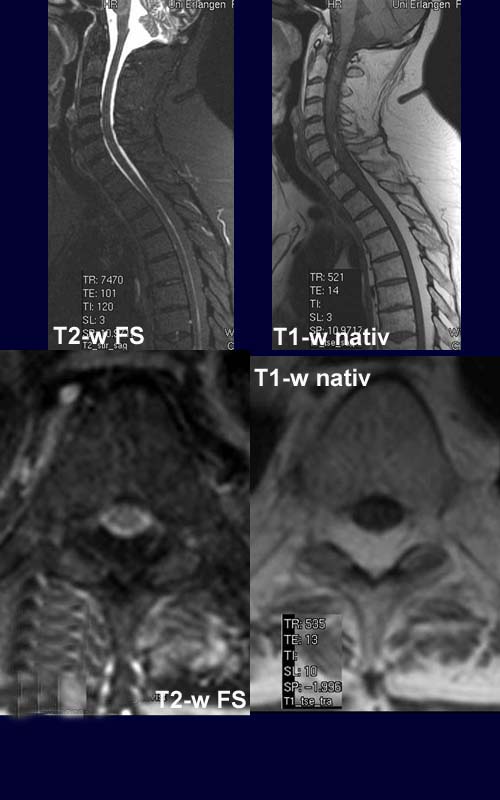
54-year-old adipose female undergoing cortison therapy.
FINDINGS:
The axial T1-w shows soft
tissue of homogeneous
high intensity in the epidural space which on sagittal T1-w can
be traced from Th1 downwards.
The T2-w fat-saturated view shows a low signal in the epidural
mass lesion as well as in the rest of the subcutaneous fat.
The postcontrast T1-w shows no
contrast enhancement.
DIAGNOSIS:
Epidural
Lipomatosis
DIFFERENTIAL
DIAGNOSIS:
Intradural Lipoma
Lipomyelocele/Lipomeningomyelocele