CASE
HISTORY/CLINIC:
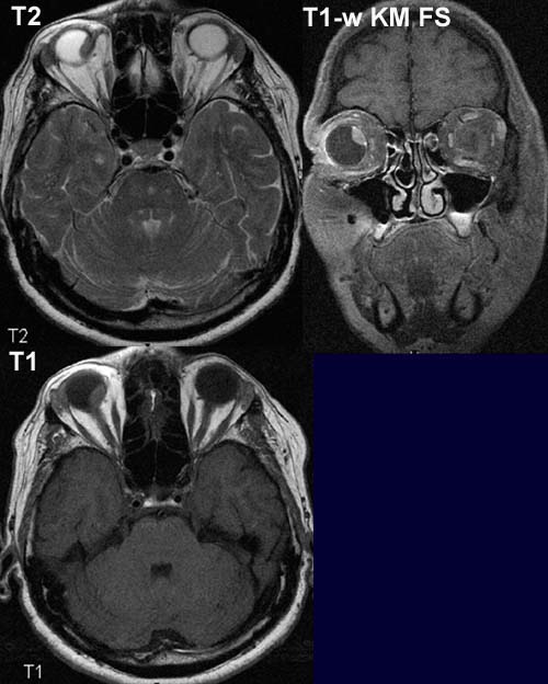
66-year-old female patient with loss of visual field on the right eye
FINDINGS:
A broad mass lesion can be seen on the inner part of the eye ball, which protrudes
into vitreous body and extends medially right up to the optic disc. The mass
lesion appears hypointense to
vitreous body on T2 and slightly hyperintense to
it on T1.
A homogeneous
contrast enhancement is seen on the postcontrast image.
DIAGNOSIS:
Uveal
melanoma on the right.
web
resources:
1) MRI-Library
2) Röntgeninstitut Nürnberg
3) Uni Graz