CASE
HISTORY/CLINIC:
45 years old Patient with bilateral Hemianopsia.
FINDINGS:
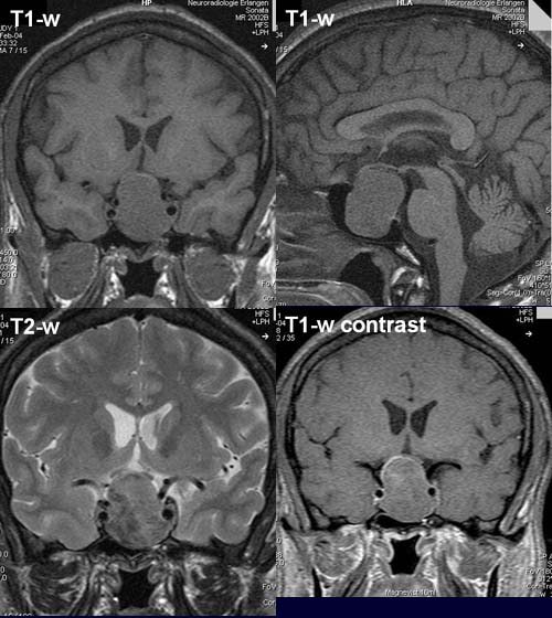
The axial and sagittal T1-w show a homogeneous
signal similar to gray matter. The T2-w image shows a well-circumscribed
intra-, supra- and parasellar mass lesion with
a somewhat inhomogeneous
signal isodense to gray matter, which compresses and displaces the optic
chiasma cranially.
Mass
lesions around the the left internal
carotid artery are more pronounced than on the right
side with mainly infiltration of the cavernous sinus.
The postcontrast axial T1-w shows a discrete peripheral
enhancement of the lesion.
The pituitary
stalk is not well defined.
DIAGNOSIS:
Pituitary
macroadenoma (> 1cm!).
DIFFERENTIAL DIAGNOSIS:
Craniopharyngeoma
Meningeoma
web resources:
1) Medizinfo
2) MRI-Library