CASE
HISTORY/CLINIC:
44-year-old patient with diagnosed metastasized melanoma.
FINDINGS:
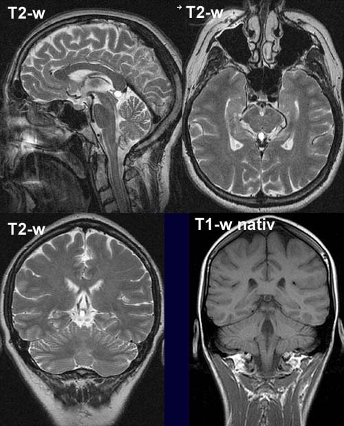
The T2-w image shows well defined hyperintense medial mass
lesion (low signal on noncontrast T1-w – not shown here) above the lamina
tecti. The T1 weighted image shows a homogeneous hypointense round
structure with a regular rim. No contrast enhancement on postcontrast T1-w image
(not shown here).
DIAGNOSIS:
No evidence of metastases.
Side
finding: Pineal
cyst.
web resources:
1) MRI-Library
2) Amersham Health