CASE
HISTORY/CLINIC:
5
years old Patient with bilateral hemianopsia.
FINDINGS:
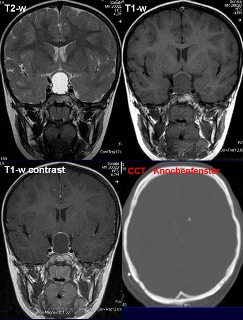
The coronary view shows a homogeneously circumscribed fluid-isointense intra-
and suprasellar
mass lesion, which compresses and displaces the optic
chiasma upwards.
The postcontrast sagittal T1-w shows a narrow peripheral rim contrast
enhancement.
The CCT (bony window) shows a shelled
calcification of the mass lesion (50-70% of the cases).
The pituitary stalk is not well circumscribed.
DIAGNOSIS:
Predominantly calcified
peripheral cystic Craniopharyngeoma.
DIFFERENTIAL DIAGNOSIS:
Pituitary adenoma (not calcified)
web
resources:
1) Medicine worldwide
2) Academicus
3) ACR Learning file
4) Scottish Radiology Society