CASE
HISTORY/CLINIC:
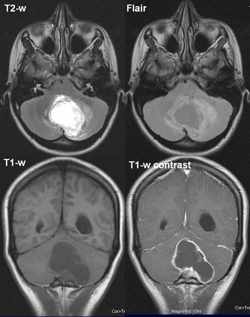
5-year-old patient with gait disturbance.
FINDINGS:
A homogeneous, almost liquor-isotense mass
lesion with an irregular periphery can be visualized in the posterior cranial
fossa. The lesion displaces the fourth ventricle ventrolaterally to the right,
leading to a subtotal compression.
Resulting enlargement of
the lateral ventricles (left > right) - see T1-w before and after contrast
administration.
The postcontrast T1-w shows a typical contrast
enhancement of the cyst wall and of the peripheral knotty portion.
The T2-w and Flair images show an inhomogeneous perifocal high
signal.
DIAGNOSIS:
Pilozytic astrocytoma with perifocal
edema and obstructive
hydrocephalus.
DIFFERENTIAL DIAGNOSIS:
Haemangioblastoma
Medulloblastoma
Metastasis
Ependymoma
web resources:
1) Medicine worldwide
2) DGN
3) ACR Learning file
4) Amersham health