CASE
HISTORY/CLINIC:
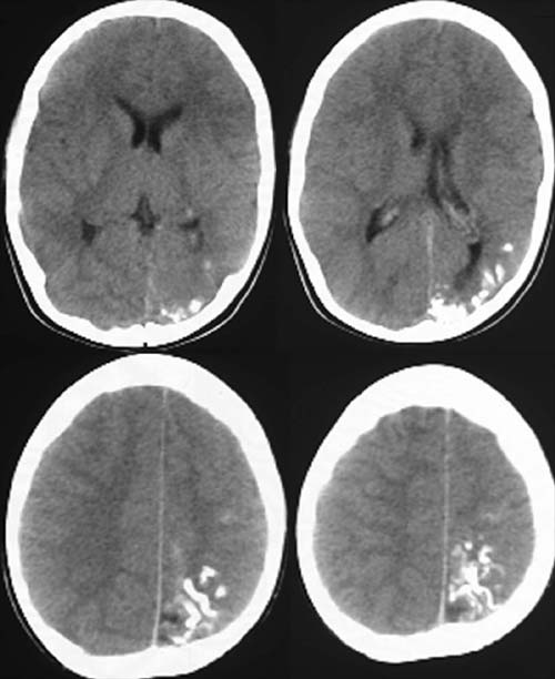
8-year-old patient with epileptic seizures. Facial nevus flammeus on the left.
FINDINGS:
Noncontrast CCT:
Evidence of hemiatrophy on
the left. Asymmetric
calvaria. Curvilinear shelled calcifications along
the left parietooccipital cerebral gyri. The calcifications
have been spreading from the occipital lobe to the frontal
lobe for 5 years.