CASE
HISTORY/CLINIC:
A 75-year-old patient without any recollection of an accident. Rapidly increasing
dementia and hemisyndrome on the right.
FINDINGS:
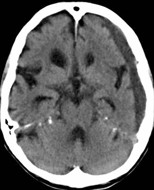
Noncontrast CCT shows a hypodense
(20-40HE) sickel-shaped mass
lesion between calvaria and brain surface on
the left. Prominently enlarged
inner and outer liquor spaces outlying
the sickel-shaped mass lesion.
DIAGNOSIS:
Chronic
subdural hematoma on the left
Side finding: cerebral
atrophy.
DIFFERENTIAL DIAGNOSIS:
Hygroma
Subdural empyema
web
resources:
1) ACR Learning file
2) MRI-Library