CASE
HISTORY:
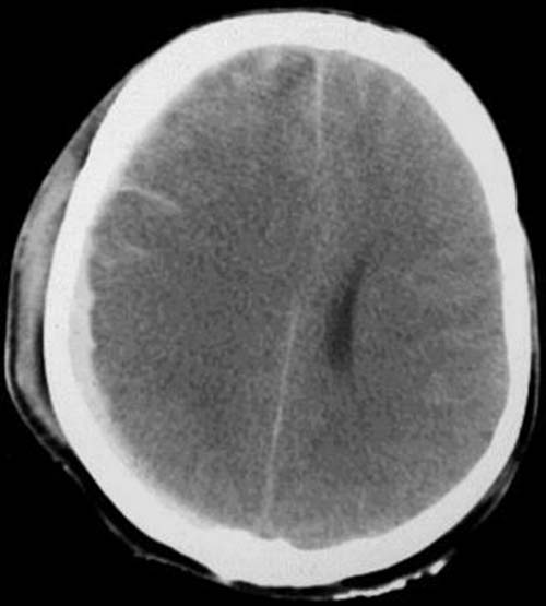
41-year-old patient after a serious head injury. Patient is unconscious.
FINDINGS:
Unenhanced CT-scan (i.e.
without contrast administration).
Hyperdense
(60-80 HE) traces of the right frontoparietal and left frontal gyri. Right-sided
parietooccipital hyperdense,
sickel-shaped mass
between skull cap and brain surface. Midline
shift to the left. Asymmetry
of the lateral ventricles. Flatenned
gyri and sulci. Hyperdense thickening
of the callus.
DIAGNOSIS:
Acute traumatic subarachnoid
hemorrhage on the right. Parietofrontal and left frontal acute
subdural hemorrhage. Right-sided frontoparietal mass with consecutive
midline
shift and compression
of the right lateral ventricle.
Brain
edema. Callus
hematoma.
DIFFERENTIAL DIAGNOSIS:
Epidural/subdural
empyema.
If
an accident is not obvious the existence of a vascular malformation (aneurysm)
must be investigated!
Information on the internet: