CASE
HISTORY:
A
71-year-old patient with a history of hypertension suddenly suffers from intense
headache and hemiplegia. Patient is comatose.
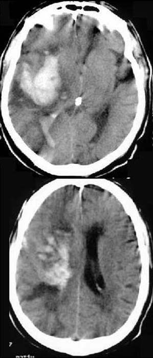
FINDINGS:
Unenhanced
CCT-scan (i.e.
without contrast administration).
Hyperdense
(60-80HE) mass on the right around the internal capsule and the basal
ganglia. Midline
shift to the left. Asymmetric
appearance
of ventricular system (compressed on the right, congested on the left). Hyperdense
collection in both the right and the left posterior horns. Flattened
gyri und sulci on the right in comparison to the left side.
DIAGNOSIS:
Hypertensive massive
hemorrhage on the right with consecutive mass
effect to the left.
Compression of the right ventricle and congestion of the left ventricle.
Bleeding
into ventricular system.
Brain
edema.
DIFFERENTIAL
DIAGNOSIS:
Brain
tumors (glioblastoma, metastases)
Hemorrhagic infarction
Cerebral abscess
Information
on the internet:
1) Uni Harvard
2) Neuroscript