CASE
HISTORY:
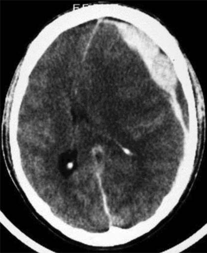
25 years old patient, unconscious after a car accident. The clinical examination
reveals a skull cap hematoma on the left.
FINDINGS:
Unenhanced
CCT (i.e.
without contrast administration).
Left frontal sickle-shaped,
hyperdense (60-80HE) mass between skull cap and brain surface. Considerable
mid-line
shift to the right. Flattened
gyri.
DIAGNOSIS:
Left-sided
acute parietofrontal
subdural
hematoma with consecutive mass effect and mid-line
shift
to the right.
Brain
edema.
Information
on the internet:
1) Uniklinik Saarland
2) PathoPic