CASE
HISTORY / CLINIC:
67-year-old patient with hoarseness and severe dysphagia.
FINDING:
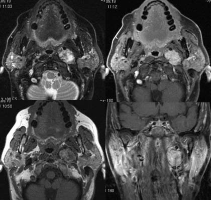
The T2-weighted
image (upper left image) demonstrates a well circumscribed inhomogeneous hyperdense mass measuring
2 cm (pepper-salt pattern). No clear distinction from the parotid
gland is seen. The T1-weighted image with neither fat suppression nor contrast
administration (lower left picture) demonstrates a hypodense, somewhat inhomogeneous mass.
After contrast administration this mass enhances
very intensely (right picture) and is generally seen to be well circunscribed.
DIAGNOSIS:
Glomus
vagale
DIFFERENTIAL
DIAGNOSIS:
Other glomus tumor
Web resources:
1) Amersham Health
2) Chorus
3) University Rochester
4) Radiosurgery
5) Emedicine
6) Theotologygroup
7) Urad.com
8) Dizzines-and-balance.com