CASE
HISTORY / CLINIC:
Female patient presenting with left-sided impaired hearing. The ear microscopic
examination revealed an effusion in the left middle ear.
The mirror examination revealed an ulcerated area
on the left side of the nasopharynx.
FINDINGS:
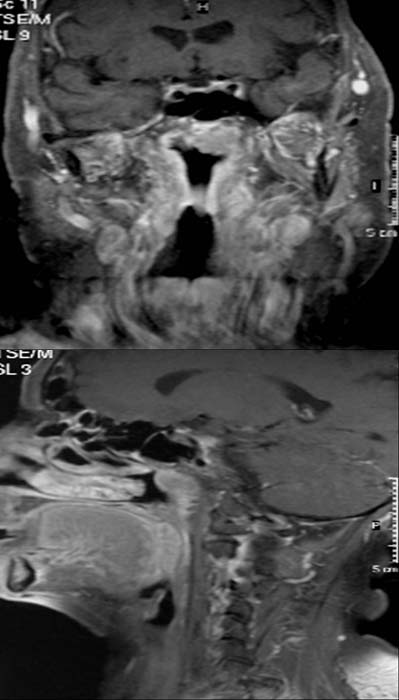
The post-contrast T1-weighted coronal image (left picture) reveals a relatively
well defined hyperdense mass in the nasopharynx that exceed the middle line.
The post-contrast T1-weighted sagittal images also demonstrate this hyperdense
mass without signs of invasion of the skull base.
DIAGNOSIS:
Nasopharyngeal carcinoma
DIFFERENTIAL DIAGNOSIS:
Lymphoma or other malignant nasopharyngeal tumor.
Web resources:
1) Amersham Health
3) Roche Lexikon Medizin
3) Medicine Worldwide
4) Pathologie Online