CASE
HISTORY / CLINIC:
47-year-old with swollen lymph nodes behind the internal jugular
vein on both sides of the neck. Recurring middle
ear effusions on the right.
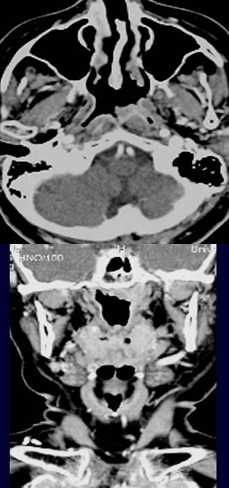
FINDINGS:
The post contrast axial CT image reveals a
small hyperdense mass at the origin of the eustachian tube. The coronal images
(lower picture) also demonstrate the same mass in
the left nasopharynx, bordering on the skull base but showing no signs of invasion.
DIAGNOSIS:
Nasopharyngeal carcinoma
DIFFERENTIAL DIAGNOSIS:
Lymphoma
Other malignant nasopharyngeal tumor.
Web resources:
1) Amersham Health
3) Roche Lexikon Medizin
3) Medicine Worldwide
4) Pathologie Online