CASE
HISTORY / CLINIC:
19-year-old young man with recurring episodes of left-sided nosebleed and hindered
nasal breathing for many years now. No other complaints.
FINDINGS:
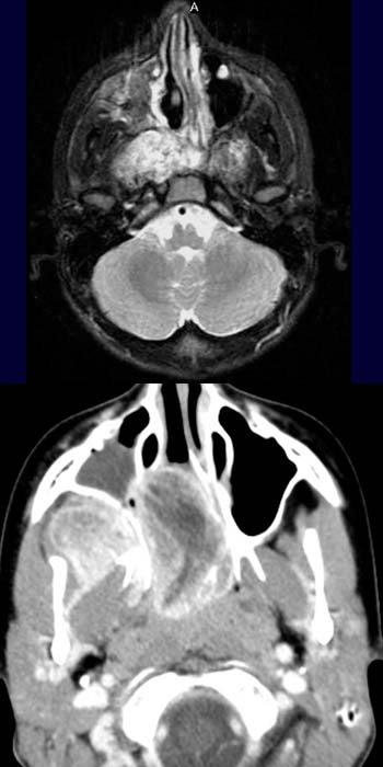
The T2-weighted image of the nasopharynx (above) demonstrates a highly
hyperdense, inhomogeneous mass in the nasopharynx as well as on both sides
of the pterygopalatine fossa. A complete
opacification of the right maxillary sinus is also demonstrated, resembling
fluid collection.
The axial CT image after contrast administration (below) also depicts this mass in
the nasopharynx and main nasal cavity. The the
spread into the right pterygopalatine fossa is also shown on this image likewise
the collection in
the right maxillary sinus.
DIAGNOSIS:
Nasopharyngeal fibroma
DIFFERENTIAL DIAGNOSIS:
Raptomyosarkoma
Nasopharyn carcinoma
Web resources: