CASE
HISTORY / CLINIC:
Patient with recurring middle ear effusions and hampered nasal breathing, occuring
intermittently for 1 year now. No B symptoms.
FINDINGS:
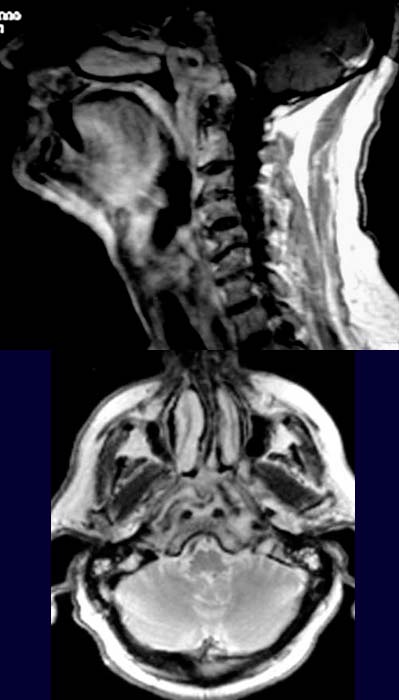
The post contrast T1-weighted sagittal image demonstrates
a hyperintense,
a bit inhomogeneous mass in the nasopharynx (above). In the T2-weighted axial
images the nasopharynx appears almost
completely radiopaque due to a hyperdense tissue (below).
DIAGNOSIS:
Hyperplastic lymphatic tissue
DIFFERENTIAL DIAGNOSIS:
Nasopharyngeal lymphoma
or carcinoma.