CASE
HISTORY / CLINIC:
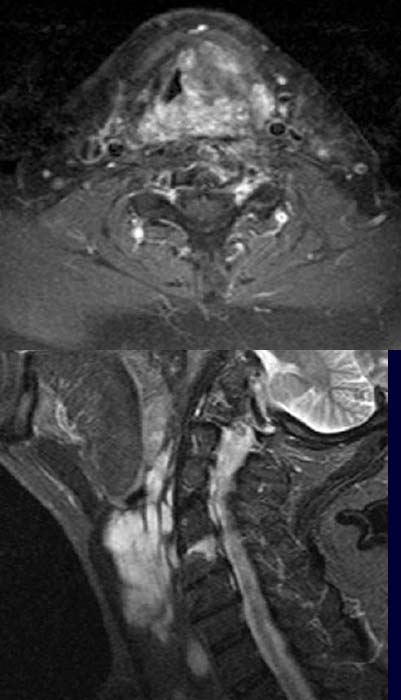
38-year-old patient with dysphagia and hoarseness.
FINDINGS:
The post contrast T1-weighted fat suppression image (above) depicts an extensive,
mainly left-sided larygeal mass with
inhomogeneous contrast enhancement.
DIAGNOSIS:
Hemangioma
DIFFERENTIAL DIAGNOSIS:
Hemangiomsarcoma
Web resources:
1) Amersham Health
2) Medecoinfo